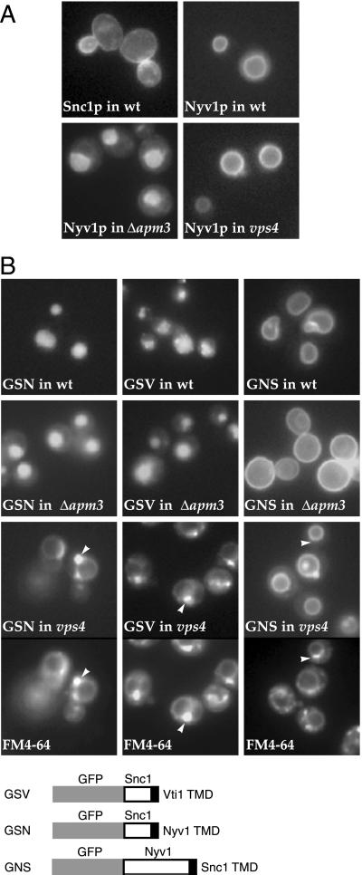
Figure 1.
Nyv1p travels to the vacuole via the AP-3 pathway. (A) GFP-tagged versions of Snc1p and Nyv1p were expressed in wild-type cells (wt), and Nyv1p was expressed in apm3 and vps4 mutants. The structures visible with GFP–Nyv1p in wild-type and vps4 cells are vacuoles. (B) GFP-tagged versions of Snc1p with the TMD from Nyv1p (GSN) or Vti1p (GSV), or GFP-tagged Nyv1p with the TMD from Snc1p (GNS), were expressed in the indicated strains. The vps4 strains were also stained with FM4-64 to reveal the PVC and vacuolar membranes; arrowheads indicate equivalent positions in the FM4-64 and GFP images.

