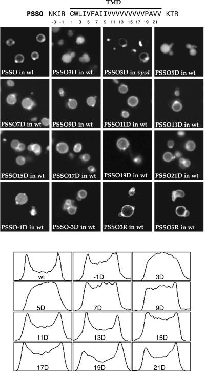
Figure 4.
Single point mutations cause internalization of PSSO. Mutants are designated by the position of the change (numbering as indicated on the sequence) and the residue at that position (D or R). Confocal images are shown for constructs expressed in wild-type cells (only vacuoles are visible), and in the case of PSSO3D, also in vps4 cells. The bottom panel shows profiles of fluorescence intensity taken across individual representative vacuoles for the Asp mutant series. The peaks visible in the wild-type (wt) profile correspond to the membrane at each side of the vacuole; intensity between these peaks indicates internal material.

