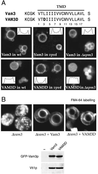
Figure 7.
Aspartic acid residues cause Vam3p to be internalized only on passage through endosomes and do not affect its function. (A) GFP-tagged Vam3p with (VAM3D) or without (Vam3) an aspartic acid at position 3 of the TMD was expressed in wild-type (wt), vps4, or apm3 cells. Confocal images of the vacuoles are shown. Insets show fluorescence profiles along a horizontal axis through the largest vacuole in each image. (B) Δvam3 cells, and cells of the same strain expressing either GFP–Vam3p or GFP–VAM3D from a plasmid, were labeled with FM4-64. Note the restoration of vacuole morphology by both proteins. The bottom panel shows immunoblots of extracts of the same cells probed for Vam3p and anti-Vam3p immunoprecipitates of these extracts probed for Vti1p.

