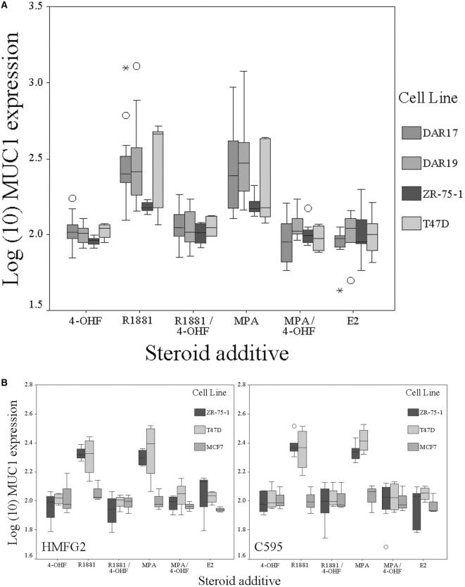
Figure 2.
Effect of exogenous steroids on membrane expression of MUC1 mucin. In each graph, log10 (change in MUC1 expression) is shown compared to control medium. (A) AR+ cell lines demonstrate a significant increase in membranous expression of MUC1 in the presence of R1881 or MPA, which is blocked in the presence of AR antagonist 4-OHF. (B) Representative results (HMFG2 and C595) obtained in AR+ (T47D and ZR-75-1) and AR- (MCF-7) breast cell lines using different antibodies to MUC1. A comparable increase of membrane MUC1 expression is seen in the presence of R1881 and MPA with different the antibodies. Expression in MCF7 is not affected by AR agonist or antagonist. In each graph, boxes represent interquartile range and error bars show confidence interval. Circles represent outlier values (1.5 to 3 boxlengths from interquartile range) and asterisks represent extreme values (greater than three boxlengths from interquartile range). For statistical analysis, see Table 3.

