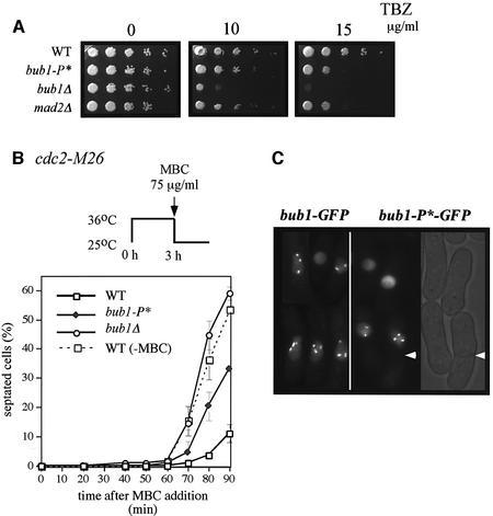
Fig. 3. Bub1-P* mutant cells are checkpoint defective. (A) WT, bub1-P*, bub1Δ and mad2Δ cells were grown to exponential phase, spotted onto YE5S plates containing 0, 10 or 15 µg/ml TBZ and incubated for 2–3 days. (B) Cdc2-M26 (WT), cdc2-M26 bub1-P* (bub1-P*), cdc2-M26 bub1Δ (bub1Δ) strains were incubated at 36°C for 3 h, released to 25°C in the presence of 75µg/ml MBC and incubated for the indicated time in minutes. Cdc2-M26 strain released in the absence of MBC (WT, –MBC) is indicated as a control. Septated cells were scored by calcofluor staining. (C) Bub1–GFPp and Bub1-P*–GFPp were observed by live fluorescence microscopy after 40 min of release at 25°C in the presence of MBC.

An official website of the United States government
Here's how you know
Official websites use .gov
A
.gov website belongs to an official
government organization in the United States.
Secure .gov websites use HTTPS
A lock (
) or https:// means you've safely
connected to the .gov website. Share sensitive
information only on official, secure websites.
