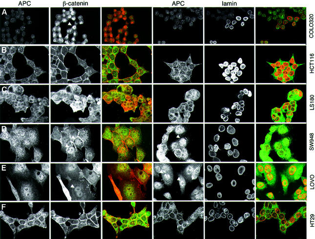
Fig. 2. Subcellular distribution of APC in colorectal cancer cell lines. Confocal sections through colorectal cancer cells from lines with a wild-type (B and C) or truncated APC (A and D–F), stained with anti-M-APC and anti-β-catenin as indicated; staining against lamin was used to mark the nuclear envelopes (merges: green, APC; red, β-catenin or lamin). Arrowheads indicate nuclear APC or β-catenin. APC is nuclear in cells with APC type I truncations (D and E), but excluded from nuclei in cells with wild-type APC (B and C) or with APC type II truncations that retain NES1506 (F). Note that the short type I truncation of COLO320 cells is not recognized by anti-M-APC (see also Figure 4B).

An official website of the United States government
Here's how you know
Official websites use .gov
A
.gov website belongs to an official
government organization in the United States.
Secure .gov websites use HTTPS
A lock (
) or https:// means you've safely
connected to the .gov website. Share sensitive
information only on official, secure websites.
