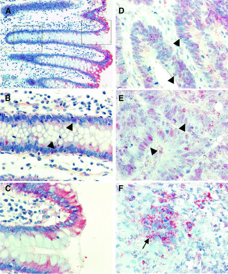
Fig. 3. Subcellular distribution of APC in colorectal carcinomas. Sections through colorectal carcinomas (D–F; two different tumours) and adjacent normal colonic mucosa (A–C), stained with anti-M-APC (red) and with hemalaun to mark the nuclei (blue); the image in (A) is taken at low magnification, to visualize the crypt axis. APC is cytoplasmic in normal cells above the crypt (A and C), but detectable in the crypt cell nuclei (B, arrowheads). In most carcinoma cells, APC is partly nuclear (D and E, arrowheads); high levels of granular cytoplasmic APC are seen in de-differentiated ‘mesenchymal’ cells at the invasive front (F, arrow).

An official website of the United States government
Here's how you know
Official websites use .gov
A
.gov website belongs to an official
government organization in the United States.
Secure .gov websites use HTTPS
A lock (
) or https:// means you've safely
connected to the .gov website. Share sensitive
information only on official, secure websites.
