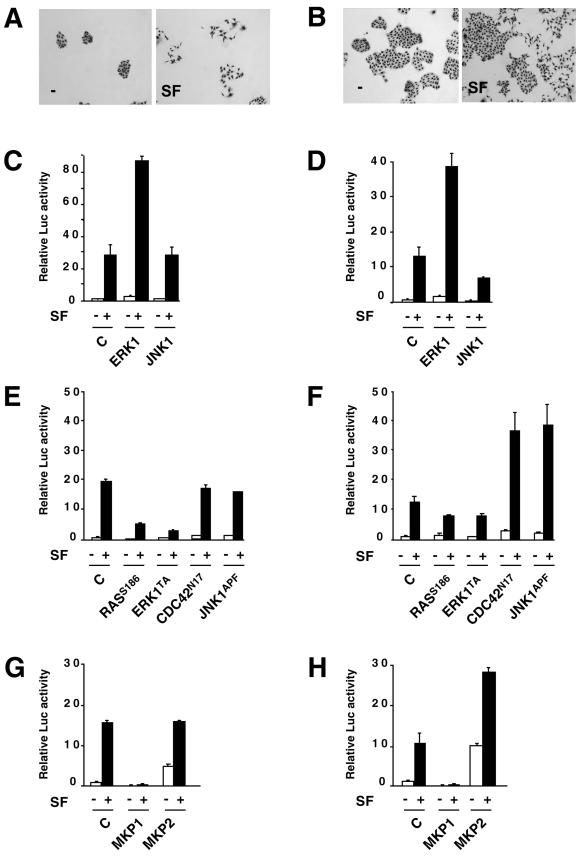
Figure 7.
Effects of ERK1, JNK1, MKP2, and MKP1 in inducing an EBS/AP1-dependent transcriptional response at different cell densities. (A and B) Scattering. Cells were seeded on 12-well plates (1250 cells [A] and 5000 cells [B]) and cultured for 2 d. The medium was replaced by DMEM–0.5% FCS, and SF/HGF (10 ng/ml) was added (SF) or not (−) for 24 h. (C and D) Effects of ERK1 and JNK1 on SF/HGF-induced transactivation of the EBS/AP1-Luc reporter vector. Cells were seeded on 12-well plates (10,000 cells [C] and 30,000 cells [D]). The next day, they were cotransfected with the EBS/AP1-Luc reporter vector and with expression vectors, either empty (C) or encoding wild-type ERK1 (ERK1) or wild-type JNK1 (JNK1). The following day, cells were left untreated (−) or treated with 10 ng/ml SF/HGF (+), and luciferase activity was measured 24 h later. The usual condition of transfection is obtained by seeding the cells at 30,000 cells per well, which gives a confluence of 50–60% at the end of the experiment. It is worth noting that at the end of the assay, the sizes of the cell islets are comparable between the cell-scattering (A and B) and transactivation (C and D) assays because of cell mortality during the transfection procedure. (E and F) Effects of dominant negative mutants of RAS, ERK1, CDC42, and JNK1 on SF/HGF-induced transactivation of the EBS/AP1-Luc reporter vector. Cells were seeded on 12-well plates (10,000 cells [E] and 30,000 cells [F]). The next day, they were cotransfected with the EBS/AP1-Luc reporter vector and with expression vectors, either empty (C) or encoding dominant negative forms of RAS (RASS186), ERK (ERK1TA), CDC42 (CDC42N17), or JNK (JNK1APF). The following day, cells were treated with SF/HGF (10 ng/ml) (black bars) or not (white bars), and luciferase activity was measured 24 h later. (G and H) Effects of MKP1 and MKP2 on SF/HGF-induced transactivation of the EBS/AP1-Luc reporter vector. Cells were seeded on 12-well plates (10,000 cells [G] and 30,000 cells [H]). The next day, they were cotransfected with the EBS/AP1-Luc reporter vector and with expression vectors, either empty (C) or encoding wild-type MKP1 (MKP1) or MKP2 (MKP2). The following day, cells were treated with SF/HGF (10 ng/ml) (black bars) or not (white bars), and luciferase activity was measured 24 h later.

