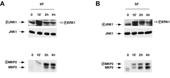
Figure 8.
Effects of SF/HGF on phosphorylation of ERK and JNK and on expression of MKP2 at different cell densities. Cells were seeded on 100-mm plates (100,000 cells [A] and 400,000 cells [B]). The next day, the cells were incubated in DMEM–0.5% FCS. The following day, cells were treated for the indicated times with SF/HGF (30 ng/ml). Cell extracts and immunoblot analysis of phosphorylated JNK1 or MKP2 expression were performed as described in Figure 5. The filters were stripped and reprobed with the anti-JNK antibody.

