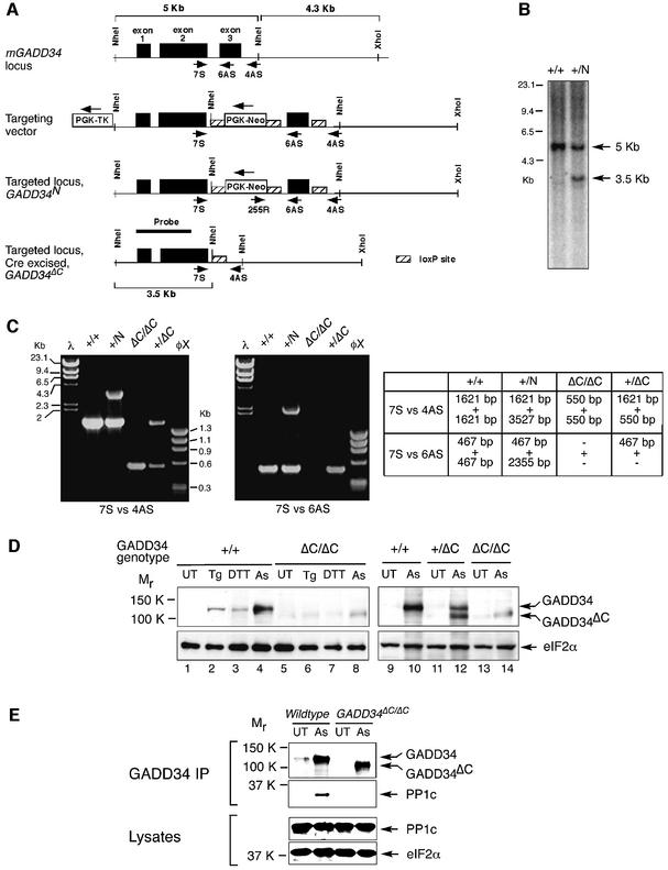
Fig. 3. Targeted mutagenesis of GADD34. (A) Scheme of the genomic organization of mouse GADD34 and targeting strategy to delete exon 3 encoding the PP1c-interacting domain (amino acid residues 549–657). From the top down: the wild-type locus; the targeting vector with the position of the oligonucleotides used to genotype the derivative alleles (arrows) and the loxP-sites (hatched rectangles) showing; the targeted GADD34N locus before Cre-mediated excision of the Neo-cassette and exon 3; and the mutant GADD34ΔC allele after Cre-mediated excision of exon 3 and the Neo cassette. (B) Southern blot analysis of NheI-digested genomic DNA of the indicated GADD34 genotypes. The position of the radiolabeled probe (HinDIII cDNA fragment containing exon 1 and part of exon 2) and the predicted genomic GADD34 NheI fragments (5 and 3.5 Kb) are indicated in (A) above. (C) Detection of the various GADD34 alleles by PCR. The primers 7S versus 4AS, and 7S versus 6AS are shown in (A). The table indicates the expected PCR products for each GADD34 genotype. +, wild-type locus; N, targeted locus; ΔC, targeted locus after CRE-mediated excised of exon 3 and the neo cassette. (D) Immunoblot analysis of GADD34 gene product in untreated (UT), thapsigargin (Tg)-, dithiothreitol (DTT)- and arsenite (As)-treated wild-type (+/+), GADD34ΔC/+ and GADD34ΔC/ΔC fibroblasts. eIF2α immunoblotting serves as a control for loading. (E) GADD34 and protein phosphatase 1 (PP1c) immunoblot of GADD34–PP1c complexes immunoprecipitated with anti-GADD34 antiserum from untreated (UT) and arsenite (As)-treated wild-type and GADD34ΔC/ΔC cells (upper panels). Immunoblot of PP1c and total eIF2α in the lysate that served as the input for the GADD34–PP1c complex immunoprecipitation (two lower panels).

An official website of the United States government
Here's how you know
Official websites use .gov
A
.gov website belongs to an official
government organization in the United States.
Secure .gov websites use HTTPS
A lock (
) or https:// means you've safely
connected to the .gov website. Share sensitive
information only on official, secure websites.
