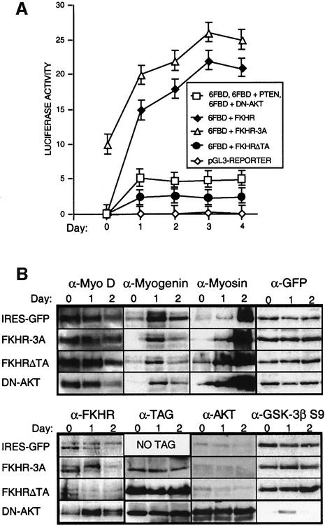
Fig. 7. FKHR activity during the differentiation of primary mouse myoblasts. (A) Luciferase reporter-based analyses of FKHR transcriptional activity of myoblasts transfected with pGL3, 6FBD, 6FBD + FKHR, 6FBD + FKHR-3A and 6FBD + FKHRΔTA during 4 days of differentiation. Co-expression of PTEN or DN-Akt produced luciferase responses that were identical to those of myoblasts transfected with 6FBD alone. Means of at least three independent experiments are given for each transfection. Error bars show the variance at each data point. (B) Western blot analyses of primary myoblasts transduced with IRES-GFP, FKHR-3A, FKHRΔTA and DN-Akt retroviral vectors, which have no effect on the levels and timing of expression of early (MyoD), intermediate (myogenin) and late (myosin) markers during muscle cell differentiation. Overexpression of DN-Akt was confirmed using an Akt antibody. The efficacy of DN-Akt in repressing known targets of Akt was assessed by determining the level of phospho-GSK-3b Ser9, which was significantly underphosphorylated in myoblasts overexpressing DN-Akt. GFP levels were used as a loading control. FKHR-3A and FKHRΔTA were detected with an antibody to the N-terminal His6 tag, and DN-Akt with an antibody to the N-terminal HA tag. The expression patterns of these markers in PTEN- and FKHR-transduced myoblasts were indistinguishable from those transduced with the IRES-GFP vector (data not shown). In all lanes, 50 µg of protein lysate was loaded, but 100 µg of FKHR-3A was required for its detection with the His6 antibody. Exposure times for the different samples detected by the same antibody were identical. The exposure time to detect FKHR-3A was 40 times longer than that to detect the TAG of FKHRΔTA and DN-Akt.

An official website of the United States government
Here's how you know
Official websites use .gov
A
.gov website belongs to an official
government organization in the United States.
Secure .gov websites use HTTPS
A lock (
) or https:// means you've safely
connected to the .gov website. Share sensitive
information only on official, secure websites.
