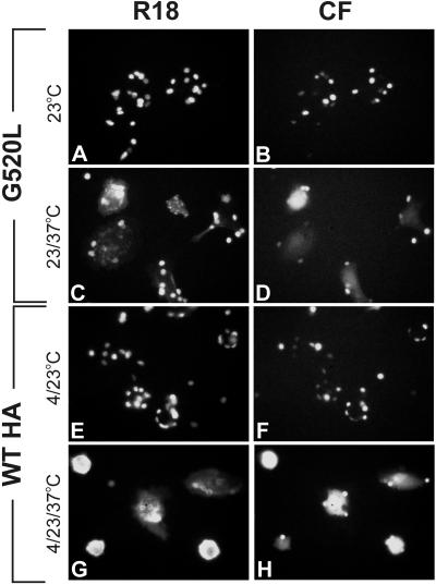
Figure 1.
Membrane (octadecylrhodamine B [R18], left column) and aqueous (CF, right column) dye redistribution between HA-expressing cells and double-labeled RBCs. (A and B) G520L cell–RBC complexes were exposed to pH 4.8 for 2 min at 23°C, followed by incubating at neutral pH at 23°C for 10 min to achieve the GL23 intermediate. Neither dye spread. (C and D) Upon increasing temperature to 37°C, both aqueous and lipid dye spread, signifying fusion. (E and F) HA cell–RBC complexes were exposed to pH 4.8 for 2 min at 4°C, followed by reneutralization at 4°C for 10 min. The temperature was then increased to 23°C (i.e., it is HA4-23). Neither lipids nor contents mixed. (G and H) The cells were subsequently incubated at 37°C for 3 min, which induced extensive fusion, as seen by the aqueous and membrane dye transfer.

