Figure 1.
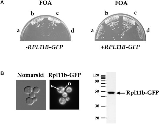
The RPL11B–GFP gene fusion is functional in yeast. (A) Four spores of a representative tetrad from the cross between PSY2083 (rpl11a::HIS3) covered by YCp50L11A URA3 CEN and PSY2084 (rpl11b::HIS3) streaked on synthetic complete medium containing FOA (left panel). Spores a and d are HIS+ and FOAS and spores b and c are his− and FOAR. Spores a–d were transformed with pPS2167 (RPL11B–GFP LEU2 CEN) and streaked on synthetic complete medium containing FOA (right panel). (B) Rpl11b–GFP expressed from a CEN plasmid in wild-type yeast was visualized by fluorescence microscopy of living cells (left panel). Arrows point to a vacuole (v) and the nucleus (n). Nomarski, phase-contrast image of cells; Rpl11b–GFP, fluorescence signal. Yeast lysate prepared from wild-type yeast cells expressing Rpl11b–GFP was probed with anti-GFP to detect Rpl11b–GFP (right panel). Five micrograms of total protein of lysate was loaded. Migration of a 10-kDa ladder of molecular mass markers is shown.

