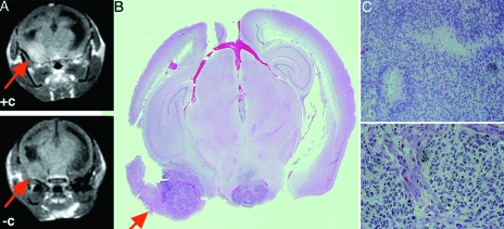
Figure 4.
Images and histopathologic sections of brain tumor obtained by infecting Ntv-a INK4a-ARF-/- mice with RCAS-PDGF. (A) Pre- and postcontrast MRI demonstrating lesion enhancement post Gd-DTPA (arrow). (B,C) Whole mount of the section shown in (A) revealing that the enhancing region has higher cellular density and necrosis.

