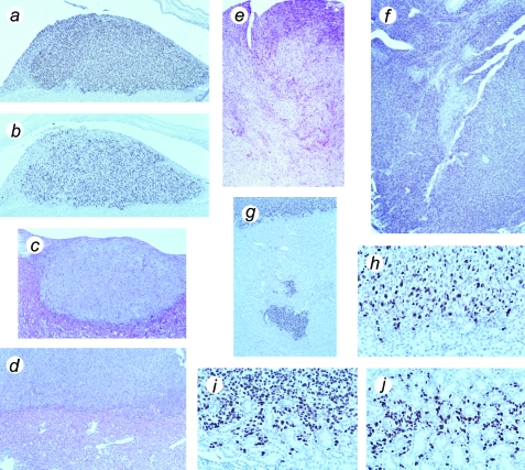
Figure 3.
Histological appearance of tumors formed from hTERT→SV40 TAg→Ras cells, showing proliferation and invasive behavior. (a) Tumor mass above mouse kidney, removed at 30 days, stained with an antibody against SV40 TAg. (b) Serial section from same specimen stained for Ki-67 proliferation-specific antigen. (c) Hematoxylin and eosin-stained section of tumor mass in relation to kidney, removed at 30 days. (d) Hematoxylin and eosin-stained section of tumor mass above kidney, removed at 60 days. (e) Hematoxylin and eosin-stained section of late tumor, removed at 75 days, showing advanced invasion and destruction of mouse kidney. (f) Similar stage of tumor stained for SV40 TAg. (g) Part of kidney at 60 days, stained for SV40 TAg, showing detached mass of tumor cells within kidney. (h) Similar specimen showing border of kidney and tumor, stained for Ki-67 antigen (note that mouse Ki-67 is not recognized by this antibody). (i,j) Similar specimen, stained for SV40 TAg, showing invasion of tumor cells into kidney; note cells surrounding kidney tubules in (j). Magnification (a–d), x100; (e–g), x50; (h–j), x400.

