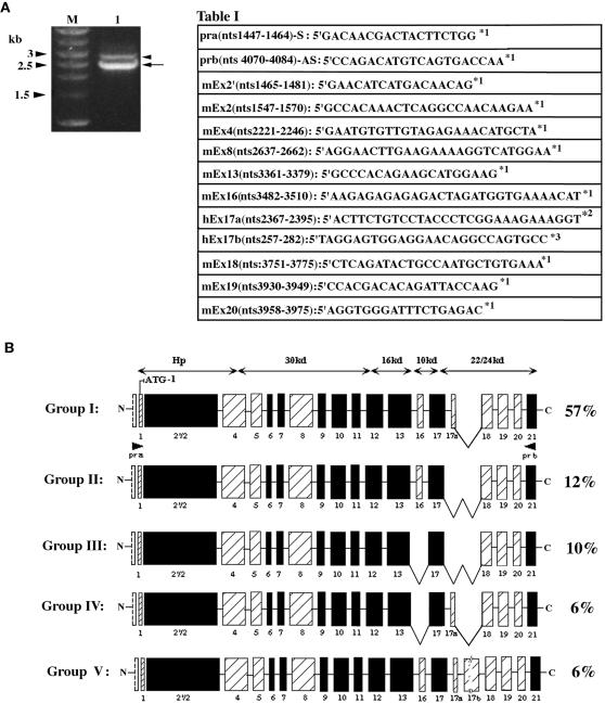
Figure 1.
Characterization of 4.1R messages in adult mouse skeletal muscle by RT-PCR analysis. (A) One-percent agarose gel electrophoresis revealed the presence of two amplification bands of ∼2.6 and ∼3 kb (arrow and arrowhead, respectively; for detailed description, see RESULTS). The table shows the nucleotide sequences of primers a and b, used in the PCR amplification assay, and the exon-specific oligonucleotide probes, used in the screening process of the skeletal muscle 4.1R cDNA library. h, human; m, mouse; nts, nucleotides; *1, GenBank accession number L00919; *2, Baklouti et al., 1997; *3, Schischmanoff et al., 1997. (B) Diagram and percentile prevalence of the major 4.1R isoforms identified in adult mouse skeletal myofibers. Previously characterized constitutive sequences are indicated as black boxes, and alternatively spliced cassettes are depicted as shaded boxes.

