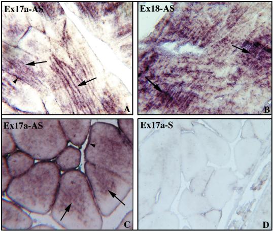
Figure 2.
In situ hybridization photomicrographs illustrating the distribution pattern of 4.1R transcripts in longitudinal sections (A, B, and D) and cross-sections (C) of skeletal muscle. Two different 4.1R oligonucleotide probes were used in the antisense orientation: E17a (A and C) and E18 (B). 4.1R mRNA is localized predominantly within the sarcoplasm (A–C, arrows), whereas an occasional staining at the periphery of skeletal fibers was also detected (A and C, arrowheads). The specific signal was eliminated completely when E17a probe was used in the sense orientation (D). Magnification, ×40.

