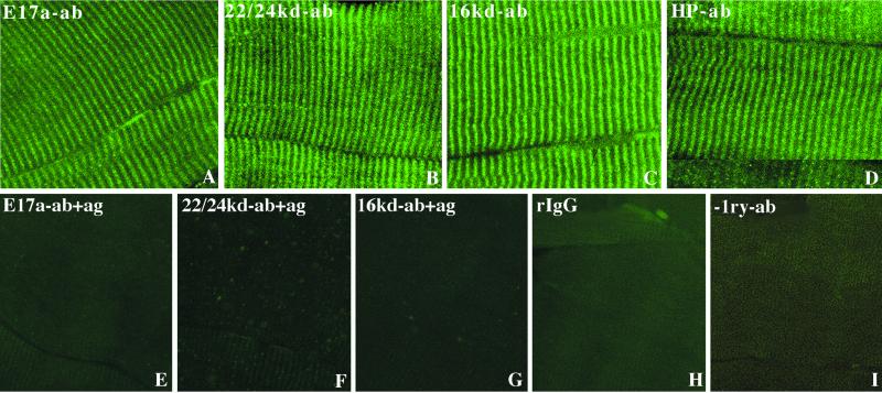
Figure 4.
Analysis of the localization pattern of 4.1R protein isoform(s) in adult skeletal muscle by immunofluorescence microscopy. A panel of four 4.1R antibodies was used with distinct domain specificities: anti-E17a (A), anti-22/24 kDa (B), anti-16 kDa (C), and anti-Hp (D). A highly ordered pattern of cross-striations was observed in all cases. In control immunodepletion experiments, anti-E17a, anti-22/24 kDa, and anti-16 kDa antibodies were preabsorbed in the presence of their respective antigens (panels E, F, and G, respectively). Furthermore, when the primary antibodies were either replaced by rabbit IgGs (H) or omitted (I), no specific signal was detected. Magnification, ×40.

