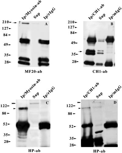
Figure 7.
Reverse coimmunoprecipitation assays of sarcomeric myosin as well as tropomyosin and protein 4.1R from total skeletal muscle lysates. An anti-myosin polyclonal antibody (A and C) and anti-tropomyosin CH1 mAb (B and D) were used in the immunoprecipitation assays, along with control rabbit (A and C) and mouse (B and D) IgGs. The immunoprecipitates were analyzed by Western blotting with anti-myosin MF20 mAb (A), anti-tropomyosin CH1 mAb (B), and anti-4.1R Hp antibody (C and D). One-sixth of the immunoprecipitates (Ip) were loaded in A and B, and one-half of the same immunoprecipitates were loaded in D. In all cases, one-eighth of the supernatant fractions (Sup) were loaded. The presence of myosin (A) and tropomyosin (B) is indicated by arrows, whereas the ∼105/110- and ∼135-kDa 4.1R isoforms are denoted by single and double arrowheads, respectively (C and D).

