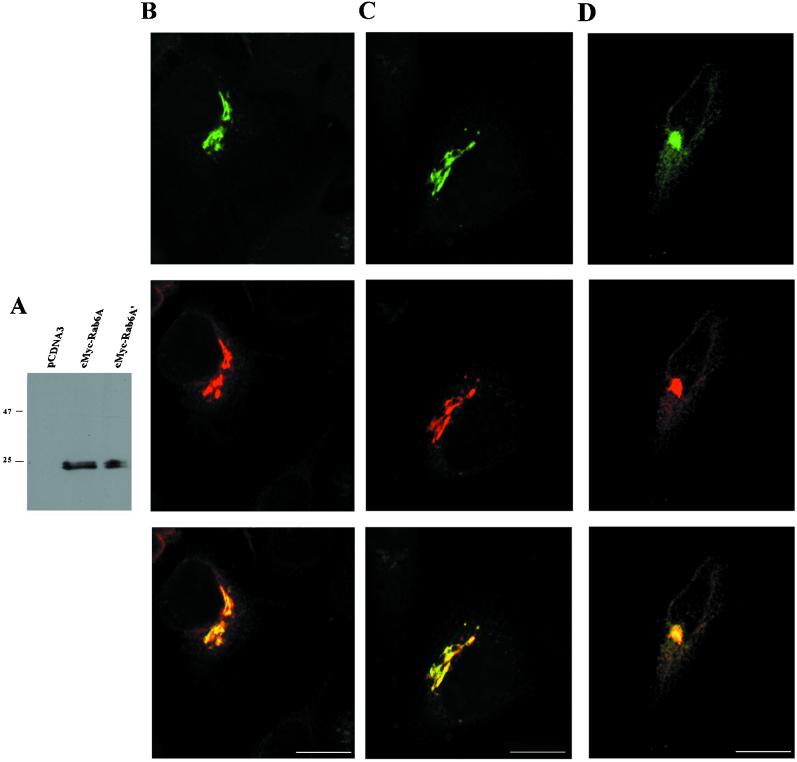
Figure 5.
Both Rab6A and Rab6A′ colocalize at the Golgi apparatus. (A) HeLa cells were transfected with empty, cMyc–Rab6A-, or cMyc–Rab6A′-encoding plasmids, and cell lysates were subjected to immunoblotting with anti-cMyc antibody. (B–D) HeLa cells were cotransfected with plasmids encoding GFP-coupled N-acetylglucosaminyltransferase I and cMyc–Rab6A (column B), GFP-coupled N-acetylglucosaminyltransferase I and cMyc–Rab6A′ (column C), or GFP-coupled Rab6A′ and cMyc–Rab6A (column D). Cells were fixed with 1% paraformaldehyde, immunostained with anti-cMyc antibodies, and viewed with a confocal microscope. Upper panels, localization of GFP fusions (green); middle panels, c-Myc staining (red); lower panels, merged images. Scale bar, 10 μm.

