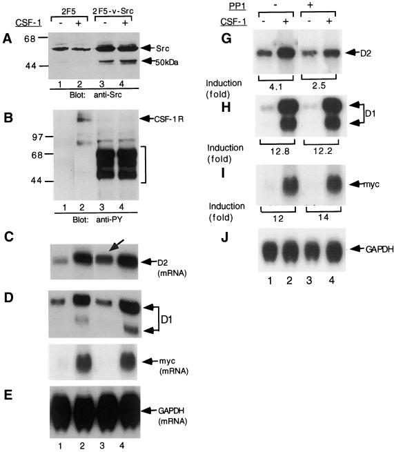
Figure 5.
v-Src enhances D2 expression and PPI inhibits D2 expression. (A) Total lysates of 2F5-v-Src cells treated with or without CSF-1 (13.2 nM, 2 min for A and B) were analyzed for v-Src protein expression by anti-Src immunoblot. (B) The same lysates were tested for enhanced protein tyrosine phosphorylation by anti-PY immunoblot. (C–F) Total RNA was isolated from cells treated with or without CSF-1 (4.2 nM, 5 h) and analyzed for increased D2 (C), D1 (D), c-myc (E), and GAPDH (F) expression. (G–J) Cells were pretreated with PP1 (1 μM) for 4 h before addition of CSF-1 (4.4 nM, 5 h), and the total RNA was analyzed for D2 (G), D1 (H), c-myc (I), and GAPDH (J) expression. The relative fold increases (measured by scanning densitometry) were based on calibration with the corresponding GAPDH levels: band+/band− × GAPDH−/GAPDH+ = Fold.

