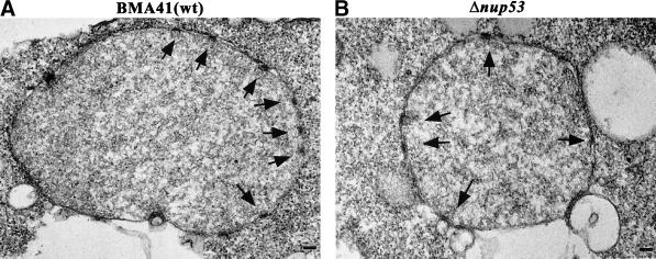
Figure 3.
Thin-section electron micrographs of Δnup53 cells (B) and of a wild-type BMA41 control strain (A) (for the cell strains see Table 1). Cells were grown at 30°C, fixed, embedded, and processed for thin-section electron microscopy. The morphology of the nucleus, the NE, and the NPCs in the Δnup53 cells appears indistinguishable from that of the wild-type cells. Arrows mark the nuclear basket of the NPCs. Scale bars, 100 nm (A and B).

