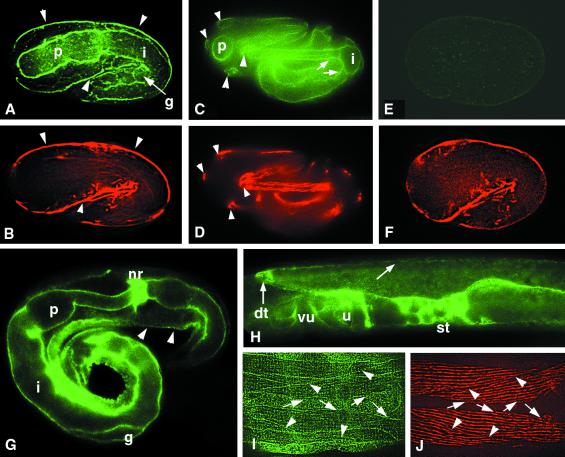
Figure 6.
NID-1 localization in cg118 and cg119 mutant animals. Animals were stained with anti–NID-1 antiserum (A, C, E, G, H, and I) or anti-myosin A (B, D, F, and J). (A and B) Twofold stage cg118 mutant embryo shows NID-1 localization under body wall muscle (arrowheads) and on the surfaces of the pharynx (p), intestine (i), and gonad (g). (C and D) Threefold stage cg118 mutant embryo showing NID-1 accumulation at the basal surfaces (arrowheads) and edges (arrows) of body wall muscle quadrants and on the surfaces of the pharynx (p) and intestine (i). (E and F) Twofold stage cg119 mutant embryo showing complete lack of staining by anti–NID-1 antibodies. (G) L1 stage cg118 mutant larva showing NID-1 accumulation at nerve ring (nr), under body wall muscle (arrow), and on the surfaces of the pharynx (p), intestine (i), and gonad (g). (H) Late L4 stage cg118 mutant larva showing NID-1 accumulation on the developing spermatheca (st), uterus (u), and vulva (vu), and at the distal tip cell (dt). Weaker accumulation on the surface of the gonad is also seen (arrow). (I and J) Ventral view of cg118 mutant adult hermaphrodite showing NID-1 accumulation at the ventral edges of the ventral body wall muscle quadrants (arrows) and at the edges between adjacent muscle cells (arrowheads).

