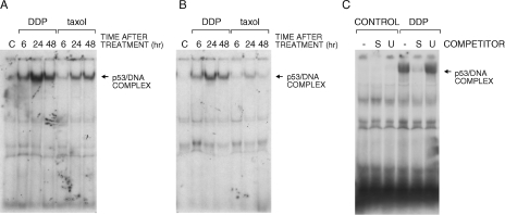
Figure 3.
Gel shift assay with extracts obtained from HCT-116 cells treated with DDP or taxol and performed with two different oligonucleotides (CON, panel A and p21, panel B), both containing a p53 binding site. Panel C reports the analysis with the CON oligonucleotide performed in the presence of a 50-fold molar excess of unlabeled specific (S) or unspecific (U) oligonucleotide.

