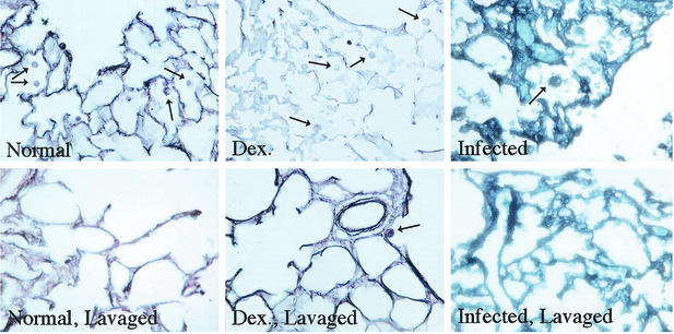
FIG. 3.
Alveolar macrophages in lung tissue before and after BAL. Lungs sections from healthy (Normal), dexamethasone-treated (Dex.), and P. carinii-infected (Infected) rats before and after lavage were assessed for alveolar macrophage number. Upper panels are sections of lungs before lavage, and lower panels are those after lavage. Arrows indicate alveolar macrophages.

