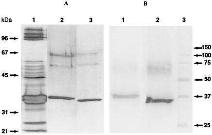
FIG. 3.
(A) SDS-polyacrylamide gel (12% polyacrylamide) under reducing conditions of the chromatographic fractions containing the leukotoxic components after staining with silver nitrate. Lane 1, culture supernatant (BHI broth) of strain 122.25; lane 2, LukM; lane 3, LukF′-PV. The two bands around 55 and 65 kDa are due to impurities in the sample buffer brought along with β-mercaptoethanol. (B) Immunoblot of culture supernatant of strain Ch122 and affinity-purified Ab to LukM (lane 1) or affinity-purified Ab to LukF′-PV (lane 2). Lane 3, molecular mass markers in kilodaltons.

