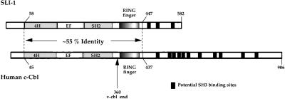
Figure 3.
Schematic alignment of SLI-1 and human c-Cbl. Conserved domains between the two proteins have been marked in the same manner. Residues 58–447 of SLI-1 share 55% amino acid sequence identity to residues 45–437 of human c-Cbl. The four-helix bundle (4H), EF hand, SH2 domain, the RING finger, and the putative SH3-binding polyproline domains are indicated. An arrow marks the C-terminal truncation point of v-Cbl. The figure is a modified version from Yoon et al. 1995 and Meng et al. 1999. The keys for this figure are retained in the structure–function analysis presentation in Figure 4.

