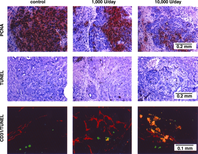
Figure 3.
Immunohistochemical analyses of livers with tumors harvested from control and IFN-α-2a-treated mice. The sections were immunostained for expression of PCNA (to show cell proliferation) and TUNEL (to show cell death). The sections were also stained with imunofluorescent anti-CD-31 antibody and TUNEL. A representative sample (x400) of this CD-31/TUNEL (fluorescent double-staining) is shown. Fluorescent red, CD-31-positive endothelial cells; fluorescent green, TUNEL-positive cells; fluorescent yellow, TUNEL-positive endothelial cells.

