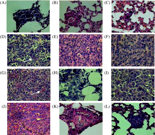
Figure 2.
High-power micrographs (x400) of 5-µm-thick histologic sections stained with hematoxylin and eosin. Sections D, E, F, G, H, and I are primary tumor sections obtained from MDA-MB-435, MDA-MB-231, MCF-7, MatLyLu, PC-3, and DU-145 tumors. Arrows indicate tumor vessels. Sections A, B, C, J, K, and L are lung sections demonstrating metastasis of MDA-MB-435 (spontaneous metastasis), MDA-MB-231 (spontaneous metastasis), MCF-7 (experimental metastasis), MatLyLu (spontaneous metastasis), PC-3 (experimental metastasis), and DU-145 (experimental metastasis) cancer cells.

