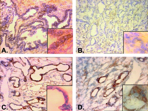
Figure 1.
Staining of normal (A, C) and malignant (B, D) prostate tissues from cases 398 (A, B) 400 (C, D) with mAb 2G8 antibody to Hsshb3p1. Staining is intense in both normal tissues shown (A, C) and in tumor tissue from case 400 (D), which was not deleted for 10p loci. Note the complete absence of staining in the malignant tissue from case 398 (B), which also demonstrated deletion of sequences adjacent to the Hsshb3p 1 locus on 10p. The large panels in (A), (B), (C), and (D) are shown at x100 magnification; insets are at x400 magnification.

