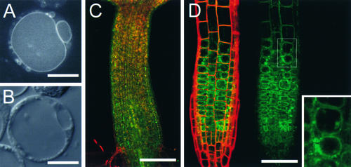
Figure 3.
Subcellular Localization of SGR2.
(A) and (B) Arabidopsis cultured cells expressing the functional SGR2-GFP fusion gene using the 35S promoter of Cauliflower mosaic virus.
(A) Confocal image.
(B) Nomarski image. GFP fluorescence was detected on the vacuolar membrane and small organelles.
(C) and (D) GFP images of sgr2-1/pSGR2::SGR2i-GFP.
SGR2-GFP fusion proteins were expressed under the native SGR2 gene promoter.
(C) The hypocotyl just after germination.
(D) The root tip.
Data from the fluorescein isothiocyanate channel (SGR2-GFP, green image) and the rhodamine channel (cell wall, red image) are merged and shown at left, and the fluorescein isothiocyanate channel data alone are shown at right. GFP fluorescence appeared to be present on tonoplasts in the plant as well as in a protoplast in (A). A magnified image of the boxed area is shown in the inset.
Bars in (A) and (B) = 10 μm; bars in (C) and (D) = 200 μm.

