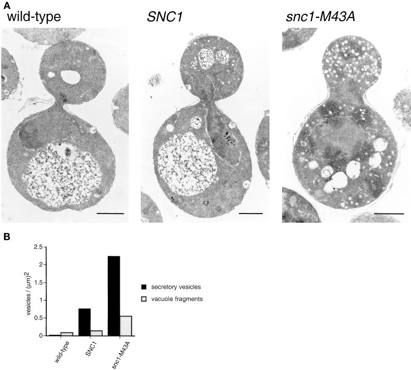
Figure 3.
Vesicle accumulation in snc1-M43A mutant yeast. (A) Electron micrographs of thin sections. Wild-type (SP1), SNC1 snc2Δ (NY2264), and snc1-M43A snc2Δ (NY2265) yeast were grown to early log phase at 25°C and then shifted to 37°C for 20 min before fixation. Bar = 1 μm. (B) Quantitation of secretory vesicle (100-nm) and fragmented (250–1000-nm) vacuole accumulation. Secretory vesicle and fragmented vacuole profiles were counted in 60 wild-type, 119 SNC1, and 75 snc1-M43A cells. The number of vesicles in each class was divided by the total surface area.

