Figure 1.
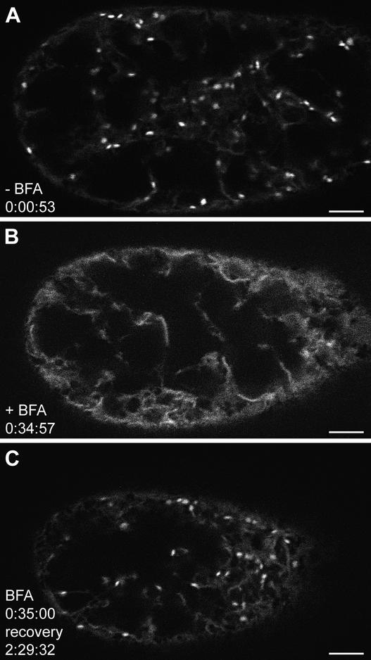
Time Lapse Confocal Observation of a Single BY-2 Cell Expressing GmMan1-GFP during BFA Treatment and Subsequent Recovery.
The cell was captured in the cortical cytoplasm close to the plasma membrane. It appears smaller at later time points because it settled slightly during the observation period. Cells were maintained in a perfusion chamber, and GFP fluorescence was captured with a confocal microscope at 34-, 64-, and 124-sec intervals as indicated (hours:minutes:seconds). See supplemental data for video at www. plantcell.org.
(A) Golgi stacks are clearly visible as bright fluorescent spots before BFA treatment. Some fluorescence also is found in a fine network, indicating the presence of a subset of the marker molecules in the ER.
(B) After 35 min in medium containing 10 μg/mL BFA, no Golgi stacks are visible. The increased ER fluorescence is caused by the relocation of the GmMan1-GFP marker into that organelle. After this time, the supply of medium is switched to fresh Murashige and Skoog medium (1962) without the toxin.
(C) During the recovery, fluorescent Golgi stacks first begin to appear after ∼60 min, although their numbers remain low until 2 hr after the removal of BFA.
Bars = 5 μm.

