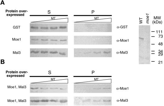
Figure 3.
Microtubules, Moe1, and Mal3 form a complex in yeast lysate. Microtubules (MT) of various concentrations (from left to right, 0, 3, 10, and 30 μM) were mixed with yeast lysates prepared from moe1Δ mal3Δ cells (strain ME1NML3A) overexpressing various proteins, as indicated on the left. The mixtures were centrifuged through a cushion buffer and both the supernatant (S) and pellet (P) were analyzed by immunoblots with antibodies indicated on the right. The anti-Moe1 antibody recognizes a single band of 62 kDa in wild-type (WT), but not moe1Δ, cell lysates (A, right). The plasmids used to express Mal3, Moe1, and GST were pREP3-MAL3, pHA-MOE1, and pAAUGST.

