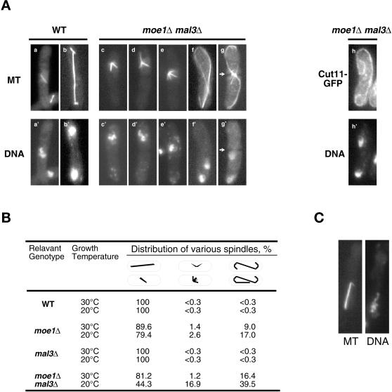
Figure 5.
Abnormal spindles in moe1Δ mal3Δ cells. Cells pregrown at 30°C to log phase were transferred to 20°C and examined after 24 h. (A) The spindles were examined by immunostaining (MT, a–g), and Cut11-GFP was visualized directly (h). An arrowhead indicates the position of a septum. (B) The relative abundance of various forms of spindles, observed in A, among all the cells that contain a spindle are tabulated. (C) An example of a moe1Δ mal3Δ cell in anaphase (spindle length of 7 μm) containing unseparated chromosomes. The strains used were SP870 (wild-type, WT), ME1UML3A (moe1Δ mal3Δ), MOE1U (moe1Δ), MAL3A (mal3Δ), and ECP16 (moe1Δ mal3Δ cut11::gfp).

