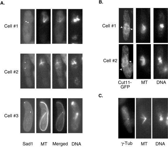
Figure 7.
Asymmetric spindle nucleation from the SPBs in moe1Δ mal3Δ cells. See legend to Figure 5 for the growth conditions of cells. (A) moe1Δ mal3Δ cells (strain ME1UML3A) were double-stained with antibodies to view Sad1 (Sad1) and the spindle (MT), and two imagines were merged by Photoshop (Adobe Systems, Inc). (B) Strain ECP16 (moe1Δ mal3Δ cut11::gfp) was double-stained with antibodies to GFP and α-tubulin to visualize Cut11-GFP and spindle (MT) simultaneously. SPBs are marked by both arrowheads and arrows; the former also mark the inactive SPBs. Note that the lower SPB in cell 2 is not on the same focal plane as the upper one; therefore, it appears smaller. (C) ME1UML3A were doubled-stained to reveal γ-tubulin (γ-Tub) and microtubules (MT). A total of 25 cells with monopolar spindles were examined, one of which is shown.

