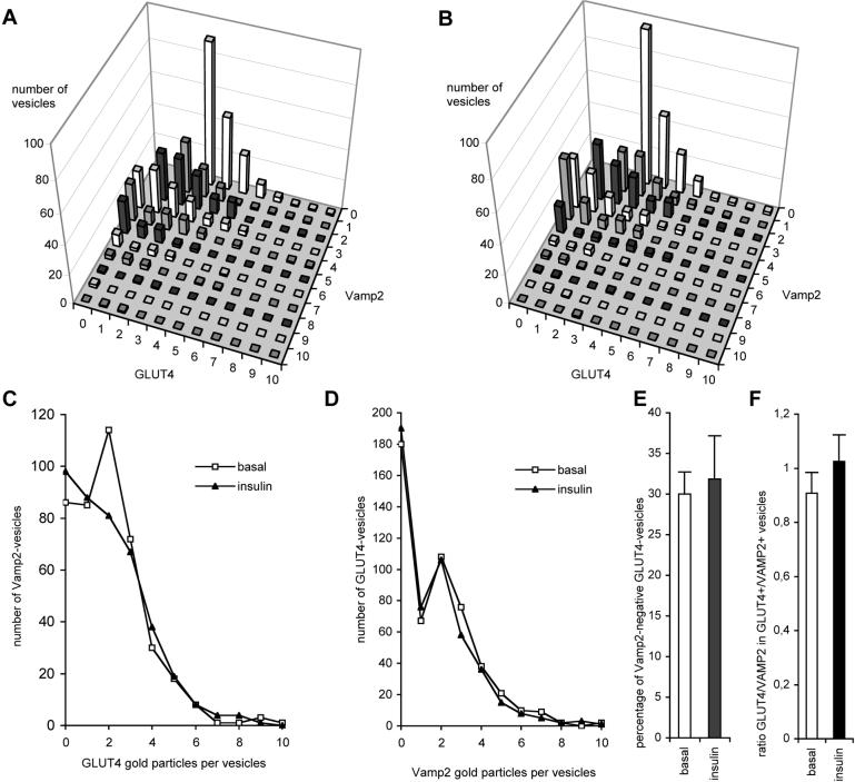
Figure 6.
Role of VAMP2 in GLUT4 translocation. Vesicles from adipocyte rims double labeled for VAMP2 and GLUT4 as shown in Figure 4, E-H were used for quantitative analysis. Counting was done in a random manner, similar to Figure 5, but now only vesicles containing ≥ 3 gold particles in total were considered. For 100 individual vesicles the number of gold particles representing VAMP2 and GLUT4 were counted (For each condition 6 × 100 vesicles were counted from two independent experiments). The distributions of all 600 counted vesicles in basal (A) or insulin treated (B) rat adipocytes are shown. Each bar represents the number of vesicles having a certain labeling characteristic. For example, the highest bar in (A) stands for 91 vesicles, each labeled with three gold particles for GLUT4 and no gold particles for VAMP2. No differences in the overall distribution of vesicles in basal (A) and insulin-stimulated (B) adipocytes were observed. Upon insulin stimulation, neither the average number of GLUT4 gold particles in VAMP2-positive vesicles (C) nor the average number of VAMP2 in GLUT4 vesicles (D) was changed. Clearly two distinct peaks representing VAMP2-negative and VAMP2-positive GLUT4-vesicles can be discerned (D). Insulin did not significantly change the percentage of VAMP2-negative GLUT4-vesicles (E). In contrast to Table 1, these quantifications include GLUT4-negative vesicles, explaining the relative lower percentage of VAMP2-negative vesicles in basal cells. The ratio of GLUT4-gold particles over VAMP2-gold particles in GLUT4-positive/VAMP2-positive vesicles remained unchanged after insulin treatment (F).

