Figure 7.
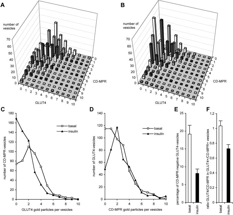
Insulin recruits GLUT4 mainly from a CD-MPR-negative GLUT4-vesicle pool, but also from CD-MPR-positive vesicles. Preparation, as shown in Figure 4D, was quantified similar to that shown in Figure 6. Compared with basal adipocytes (A) and after insulin stimulation (B), a clear change in the distribution of vesicles is visible. The average number of GLUT4 gold particles in CD-MPR-positive vesicles decreased upon insulin stimulation (C), and GLUT4 disappeared from the CD-MPR-negative vesicle pool (D). The number of vesicles containing only GLUT4 and no CD-MPR decreased upon insulin treatment (E, p < 0.005). Insulin caused a decrease of the ratio of GLUT4-gold particles over CD-MPR-gold particles in GLUT4-positive/CD-MPR-positive vesicles (F, p < 0.01).

