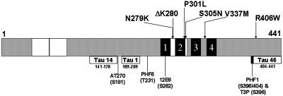
Figure 1.
Topography of FTDP-17 missense mutations in tau- and epitope-specific anti-tau antibodies. Schematic representation of the longest tau isoform, designated Tau40, containing 441 amino acids. The locations of six FTDP-17 missense mutations studied here are identified in the schematic diagram of tau. The two 29-amino-acid-long inserts are defined by white boxes at the amino-terminal region and the MT binding repeats appear as four black blocks near the carboxy terminal of tau. Each MT repeat is numbered 1–4. The defined epitopes recognized by each of the anti-tau antibodies used in this study are shown below the tau schematic with their corresponding amino acid length and position as well as the code name of the specific antibody that recognizes each epitope. The exact site(s) of phosphorylation detected by the phosphorylation-dependent anti-tau MAbs are shown here below the code names.

