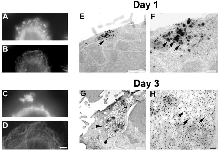
Figure 8.
Aggregates of FTDP-17 tau mutants grow larger with time. CHO cells stably expressing Wt tau and mutant tau protein were plated at low density on coverslips and grown for either 1 or 3 d. (A–D) Double-labeled immunofluorescence images of CHO cells expressing the VPR tau mutants immunostained with rabbit antirecombinant tau (17026; A and C) and a MAb to α-tubulin (B and D) in cells grown for 1 d (A and B) or 3 d (C and D). (E–H) Immuno-EM detection of tau by using 17026 as the primary antibody visualized either with secondary antibody and silver-enhanced DAB (E and F) or nanogold conjugated secondary antibody (G and H). Bar, 10 μm at 60× for immunofluorescence photomicrographs; bar, 100 nm for immuno-EM.

