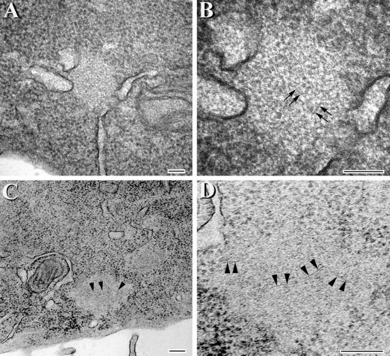
Figure 9.
Aggregates with fibrillar structures are detected in CHO cells expressing the VPR tau mutants. Transmission EM of tau aggregates at low (A and C) and corresponding high (B and D) magnifications. (C and D) Images of aggregates that were treated with formic acid. The image in D is a higher magnification of the lower left portion of the inclusion seen in C. Arrowheads in C correspond to the same region that is marked by arrowheads at higher magnification in D. Arrows in B and D highlight fibril-like structures within the aggregates. Bar, 100 nm.

