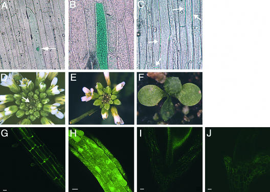
Figure 1.
CLV3 Protein Is Localized to the Apoplast.
(A) to (C) Phase-contrast optics used to detect GUS.
(A) Cell transiently expressing the GFP/GUS protein alone, which is confined to the nucleus (arrow).
(B) Cells transiently expressing the CLV3Δsp-GFP/GUS fusion protein, which is present throughout the cytoplasm.
(C) Cells transiently expressing the CLV3-GFP/GUS fusion protein, which is detected only between cells (arrows).
(D) Inflorescence apex of a clv3-2 mutant plant showing massive meristem enlargement (arrow) and production of supernumerary flowers and floral organs.
(E) Inflorescence apex of a clv3-2 mutant plant stably expressing the 35S::CLV3-G fusion protein. This transgenic T2 plant resembles the wild type, indicating that CLV3 function has been restored by the transgene.
(F) Seedling with a prematurely terminated meristem from a 35S::CLV3-G clv3-2 line. The shoot apical meristem has formed a terminal leaf.
(G) GFP fluorescence in a root from a 35S::CLV3-G wild-type T2 transgenic plant.
(H) GFP fluorescence in a root from a 35S::CLV3Δsp-G wild-type T2 transgenic plant.
(I) Autofluorescence in the cell wall of an untransformed wild-type hypocotyl.
(J) GFP fluorescence in the hypocotyl of a 35S::CLV3-G wild-type T2 transgenic plant.
Bars = 20 μm in (G) and (H) and 50 μm in (I) and (J).

