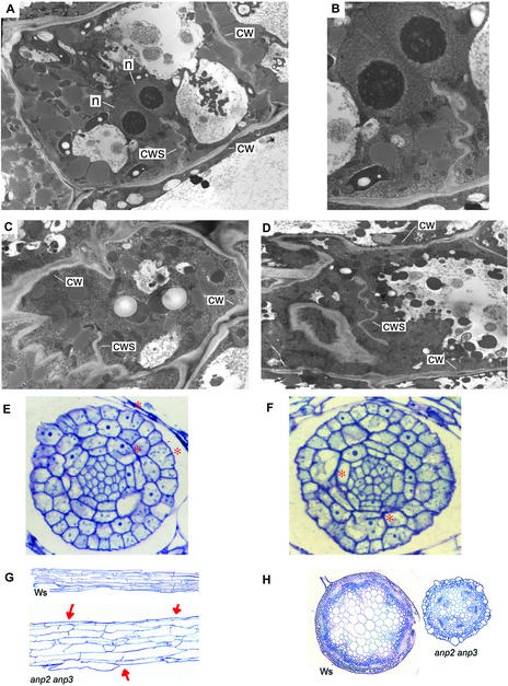
Figure 5.
Cell Division Is Disrupted in anp2 anp3 Plants.
(A) to (D) Transmission electron microscopy of hypocotyl cells of 2-day-old dark-grown anp2 anp3 plants. cw, cell wall; cws, cell wall stub; n, nucleus.
(A) Cell with two nuclei and a cell wall stub.
(B) Higher magnification view of (A).
(C) Cell with a cell wall stub.
(D) Incomplete cell wall in the middle of a cell.
(E) and (F) Longitudinal sections of anp2 anp3 embryos. Red asterisks indicate the locations of cell wall stubs. The lower asterisk in (F) indicates a binucleate cell.
(G) Longitudinal sections of 3-day-old dark-grown hypocotyls. Both sections are shown at the same magnification. Arrows indicate cell wall stubs.
(H) Transverse sections through inflorescence bolts. Both sections are shown at the same magnification.

