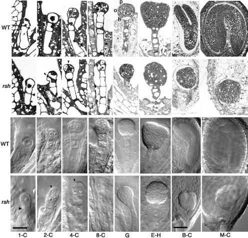
Figure 2.
Morphology of rsh/rsh Mutant Embryos Compared with Wild-Type Embryos during Development.
Stained sections (rows 1 and 2) and Nomarski images (rows 3 and 4) of wild-type (rows 1 and 3) and rsh mutant (rows 2 and 4) embryos at comparable stages of development (columns). Column 1-C, one-cell stage; column 2-C, two-cell stage; column 4-C, four-cell stage; column 8-C, eight-cell stage; column G, globule stage; column E-H, early-heart stage; column B-C, bent-cotyledon stage; column M-C, mature-cotyledon stage. Note the large apical cell in 1-C rsh, the abnormal cell shapes in 2-C and 4-C rsh (arrowheads show the dividing line in rsh), the absence of normal protoderm (p), O-ring (o), and hypophysis (h) in G rsh, and the small abnormal embryo within the normal seed coat in M-C rsh. Bar in 1-C = 20 μm for 1-C to E-H; bar in B-C = 80 μm for B-C and M-C. WT, wild type.

