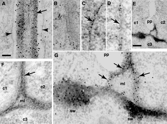
Figure 7.
Electron Micrographs of RSH-GFP Localization.
(A) Localization of RSH-GFP to the cell wall of a 3-day-old root section. Note the cell wall (arrow) between two cells with a concentration of immunogold particles (sharp black spots). Arrowheads denote the plasma membrane.
(B) Golgi and trans-Golgi network in a 3-day-old root section. Note the concentration of immunogold particles.
(C) and (D) Localization of RSH-GFP to the cell walls of heart-stage embryo cells. Note the immunogold particles in the thin walls (arrows) of young, rapidly dividing cells.
(E) Fusion of the cell plate to the cell wall in a dividing heart-stage embryo cell. Two new cells (c1 and c2) with a cell plate (pp) between them. c3 indicates an adjacent cell. Note the cell wall connections between the edge of the cell plate and the swollen regions of the mother cell wall.
(F) Localization control. This embryo section was prepared like the other sections except that preimmune serum was substituted for the first antibody (anti–RSH-GFP antibody). Note the absence of immunogold particles in the three cells (c1, c2, and c3), the cell wall (arrow), and the middle lamella (ml).
(G) Enlargement of (E). Localization of RSH-GFP as seen by the concentration of immunogold particles in swollen regions (sw) of the mother wall and new connecting walls (arrows).
Bar in (A) = 100 μm for (A) to (D), (F), and (G). Bar in (E) = 25 μm.

