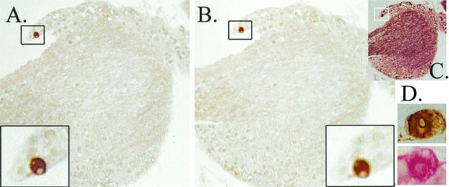
FIG. 4.
Analysis of penetration of IHC reagents into the TG. TG were first assayed for viral proteins by using WGIHC. These ganglia were then embedded in paraffin, serially sectioned, and restained by using the anti-HSV antibody and a red chromagenic substrate as detailed in Materials and Methods. (A) Portion of a latently infected TG at 22 h post-HS containing a single viral-protein-positive neuron (box). This neuron was detected during WGIHC. (B) After restaining, no additional viral protein staining was detected. (C) The same section as in panel B after being stained with a neurofilament antibody and VIP (Vector) as the chromagenic substrate, confirming that processing had not destroyed antigenicity of the tissue. (D) Brown (diaminobenzidine)- and red (Fast Red)-stained neurons.

