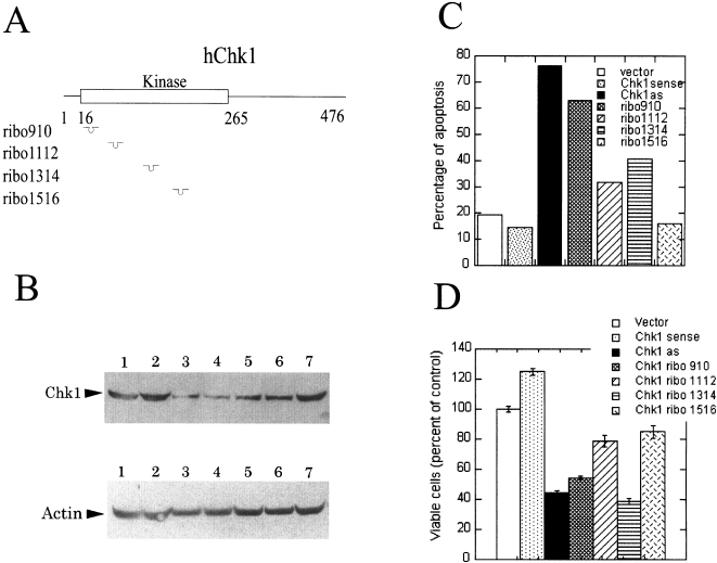
Figure 2.
Blocking Chk1 expression induces cell death. (A) Cartoon of human Chk1 gene and the hybridization positions for different ribozymes. (B) Chk1 protein levels in the transfected cells. H1299 cells were transfected with different plasmids as indicated below using LipofectAMINE PLUS. Western analysis of the total cell lysates was performed as described. The filter was blotted with anti-Chk1 antibody (top panel followed by blotting with anti-actin antibody (lower panel. Lane 1: vector control; lane 2: full-length Chk1 sense cDNA; lane 3: full-length Chk1 antisense cDNA; lane 4: ribozyme 910; lane 5: ribozyme 1112; lane 6; ribozyme 1314; lane 7: ribozyme 1516. (C) Blocking Chk1 expression induces cell death. Cells were harvested 24 hours after transfection. The apoptosis assay was carried out as described. (D) Blocking Chk1 expression decreases cell survival. H1299 cells were transfected with different plasmids as indicated. An Alamar Blue assay was carried out to determine the cell survival.

