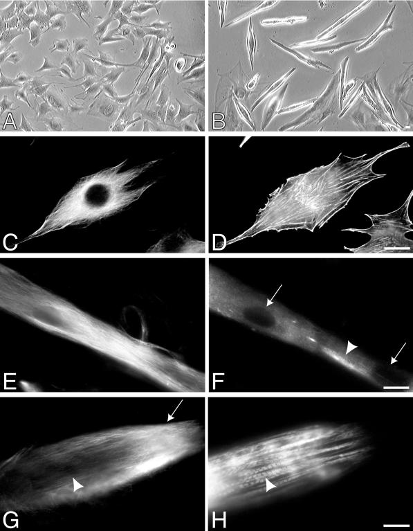
Figure 1.
Phase-contrast and immunofluorescence microscopy of differentiating mouse myoblasts (C2C12). A phase-contrast image of a field of proliferating myoblasts (A) and multinucleated myotubes (B). (C and D) Immunofluorescence microscopy of a C2C12 myoblast that has been double labeled with an antibody to tubulin (C) and FITC-conjugated phalloidin (D). (E–H) Immunofluorescence microscopy of C2C12 myofibrils that have been double labeled with antibodies to tubulin (E and G) and skeletal muscle myosin (F and H). The myotube in E and F has been differentiating for 3 d and has begun to express skeletal muscle myosin (arrowhead in F). Two visible nuclei are indicated (arrows in F). The myotube in G and H has been differentiating for 6 d and contains striated myofibrils (arrowhead in H). Note regions of decreased microtubule density (arrowhead in G) versus abundant microtubules (arrow in G) at the tip of the myofibrils. Bar, 20 μm.

