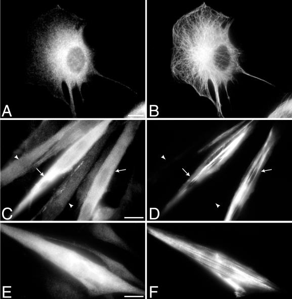
Figure 3.
Cellular localization of KIF3B in proliferating C2C12 myoblasts and differentiating C2C12 myotubes. (A and B) C2C12 myoblast labeled with α-KIF3B-T (A) and an antibody to tubulin (B). (C–F) C2C12 myotubes labeled with α-KIF3B-T (C and E) and an antibody to skeletal muscle myosin (D and F). Myotubes, 3 d (C and D); myotubes, 6 d (E and F). C2C12 myotubes differentiate at different rates; therefore, a subset is expressing and assembling skeletal muscle myosin (arrows in C and D), while others are not (arrowheads in C and D). Expression of KIF3B is brighter in cells containing skeletal muscle myosin. Bar, 20 μm.

