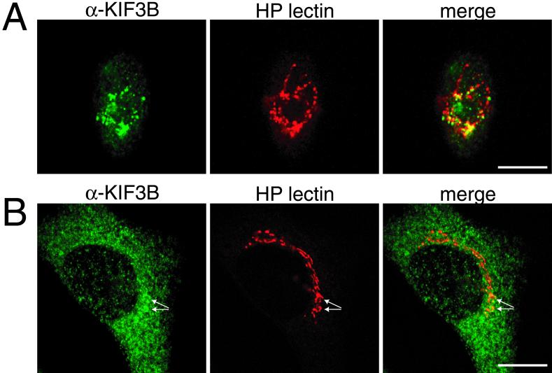
Figure 5.
KIF3B primarily colocalizes with a Golgi marker in Xenopus A6 cells. In C2C12 cells, only a small subset of KIF3B colocalizes with the same Golgi marker. (A and B) Confocal images of a Xenopus A6 cell (A) and C2C12 myoblast (B) labeled with α-KIF3B-T (green) and Texas Red-conjugated HP lectin (red), with merged images to the right. HP lectin labels the Golgi apparatus (red) in both cell types. In A6 cells, labeling of our anti-KIF3B-T antibody overlaps with HP lectin (A, yellow in merged image). In C2C12 myoblasts (B), KIF3B is diffuse and punctate throughout the cytoplasm (B, green). KIF3B does not significantly colocalize with HP lectin staining in these cells; however, there are minor aggregates of KIF3B that appear to colocalize with regions of HP lectin staining (arrows in B). Bar, 20 μm.

